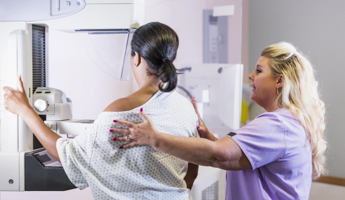
“Mammograms save lives and should be part of routine health screenings for women,” said Jana L. Deitch, MD, FACS, Director of the Breast Health Program at St. Catherine of Siena Hospital. “The new guidelines by the U.S. Preventive Services Task Force recommend women begin screening mammograms at age 40. Women should be discussing their breast health with their gynecologist. Some patients, especially those considered high risk, may need additional screenings.”
What is a mammogram?
A manual breast exam cannot always detect abnormal changes in your breasts. For that, you will need a mammogram and possibly other advanced imaging.
Mammograms are low-dose X-rays of your breasts. They allow your physician to identify abnormal changes in your breast tissue that cannot be felt during an external manual exam. When combined with a clinical breast exam done by your physician, a mammogram is the most effective way to identify breast cancer in its earliest stages. The earlier breast cancer is found, the better your chance of a successful outcome.
- Screening mammograms are used when you do not have signs of breast cancer.
- Diagnostic mammograms are used after a lump is detected or you experience other warning signs such as breast pain, discharge or changes in shape or size of your breasts.
What do the new mammogram guidelines mean for you?
Mammograms are essential to preventive care, but you may need clarification about when you should schedule your first mammogram and how often you should have one. Recently, the U.S. Preventive Services Task Force (USPSTF) has recommended all women start routine breast cancer screening at 40. The previous recommendation was by the age of 50.
USPSTF noted that this new recommendation could save 19% more lives. For women in the United States, breast cancer is the second most common cancer and the second most common cause of cancer death. Black women are 40% more likely to die of breast cancer than white women.
The new USPSTF guidelines are for women at average risk, not those at high risk due to factors like genetic history or previous breast cancer.
USPSTF recommended guidelines for mammograms
- All women should get screened for breast cancer every other year starting at age 40.
- For women with dense breasts: More research about additional breast ultrasound or MRI screening is needed.
- For women older than 75: More research about the benefits and harms of screening is needed.
Who is considered at average risk for breast cancer?
You are considered at average risk if you:
- Do not have a genetic mutation such as the BRCA gene known to increase the risk of breast cancer.
- Have not had chest radiation therapy before turning 30.
- Have no personal or strong family history of breast cancer. A strong family history means you have one or more first-degree relatives—your mom, sister or daughter—diagnosed with breast cancer.
Who is considered at high risk for breast cancer?
You are considered at high risk if you:
- Had radiation therapy between the ages of 10 and 30 years old.
- Have a confirmed BRCA1 or BRCA2 gene mutation based on testing.
- Have a first-degree relative with a BRCA1 or BRCA2 gene mutation but have not had genetic testing yourself.
- Have Cowden syndrome, Li-Fraumeni syndrome or Bannayan-Riley-Ruvalcaba syndrome or have one or more first-degree relatives with one of these syndromes.
Learn more about genetic counseling for cancer.
How often should I get screened?
The American Cancer Society (ACS) recommends annual mammograms.
Your gynecologist will discuss the best option for you based on your age and medical history.
What advanced imaging technology is used for breast cancer screening?
Advanced technology makes it possible to obtain more detailed images of your breasts.
- Breast Magnetic Resonance Imaging (MRI) uses magnets and radio waves to create detailed 3D images of your breast tissue.
- Breast ultrasound uses high-frequency sound waves for a non-invasive view of your breast. It is an effective way to find cancer in women with dense breasts and may eliminate the need for a biopsy in some cases.
- 3D mammography uses low-dose X-rays to produce computer-generated images of your breast tissues. It provides detailed, clear images to reveal cancers that would otherwise be too small to see.
- Molecular breast imaging uses a special camera and radioactive tracer to identify cancer cells. Your physician may recommend molecular breast imaging if you have dense breasts.
Schedule your screening at Catholic Health
Scheduling a mammogram can save your life. Talk to your gynecologist about the new recommendations and when a mammogram is right for you.
Catholic Health offers convenient locations in Nassau and Suffolk counties.
- Good Samaritan University Hospital Women's Imaging Center (West Islip, NY)
- Bishop McGann Women’s Imaging Center at Mercy Hospital (Rockville Centre, NY)
- St. Catherine of Siena Diagnostic Imaging (Smithtown, NY)
- Siena Women’s Health Outpatient Diagnostic Pavilion (Smithtown, NY)
- St. Charles Hospital Radiology (Port Jefferson, NY)
- The Women’s Health Center of St. Francis Hospital & Heart Center® (Roslyn, NY)
Call 866-MY-LI-DOC (866-695-4362) to find a Catholic Health physician near you.


